Fundus Camera
The surgeons at the Nazar Eye Hospital have a lot of practice performing assessments with a Fundus camera.
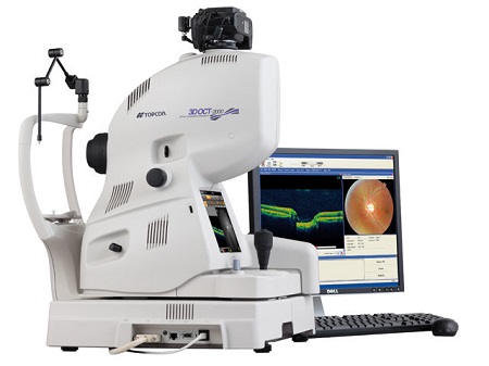
A fundus camera is basically a low power microscope with a camera attached to it. With a fundus camera,
diagnosis of the progression of diseases, diagnosis of diseases, screening and epidemiology is done in
the interior surface of the eye.
Optics Of Fundus Camera
Light is generated from the electronic flash and is sent through a set of filters and then onto a mirror
which is round. Next, this mirror reflects light into a combination of lenses focusing the light. A mask
is used on uppermost lens which gives shape to the light into a dough nut. Then, the dough nut shaped
light is reflected onto a round mirror with central aperture, exits camera through objective lens, and
goes on into the eye through cornea.
The retinal image goes out of the cornea through the un – illuminated, central part of the doughnut. Then,
the light goes on through the central aperture of the round mirror, through correction device and compensation
lenses, and back to the single lens camera system.
Fundus Retinal Photographs
Fundus photographs present the present opthalmoscopic scene of the patient’s retina. The photograph is of a
lot of value to the physician who can study it to find retinal changes.
The state of glaucoma can be assessed by fundus photography and then the physician can suggest the appropriate
therapy required for its treatment.
Diabetic retinopathy like micro aneurysms and muscular edema can can be done through fundus photographs.
It can also be used for fluorescein angiography.
The Nazar Eye Hospital is equipped with all the Fundus camera technologies. Moreover, the physicians and
ophthalmic photographers are highly skilled in their areas to asses all the information about the retina with
the help of fundus cameras.
All in all, Fundus Camera does wonders being a low power microscope with attached camera. Our surgeons get
the most out of it and the retina can easily be studied.